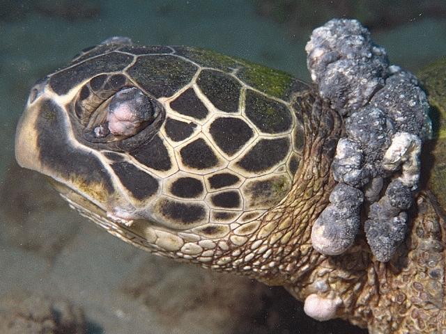
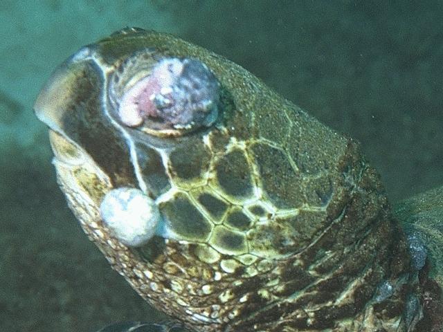
 Left side, 1992 Turtle 22: 4 Spot (68K JPEG)
We offer these images to anyone who can make use of them. We hope that someone can shed more light on what exactly these photos reveal about the eyes of these turtles.
These images were originally taken underwater at Honokowai, Maui, during the summers of 1994 and 1995.
The icons below are linked to 640x480 JPEGs. If you prefer to review the images as 240x180 GIFs, use our contact sheet (6 GIFs, 140K).
The nature of this medium makes it impossible for us to know exactly how our images will appear on other monitors. Colours can be shifted or missing entirely. The brightness, contrast, colour balance, and gamma of our monitors can vary significantly from yours. This means that you must not rely on the colours contained in these images, even if you are using a video system that has millions of colours.
Left side, 1992 Turtle 22: 4 Spot (68K JPEG)4 Spot was a clean juvenile upon arrival at the Turtle House in 1992. In 1995, this turtle has double tumors covering the left eye, a small tumor protruding from the left side of the mouth, and numerous tumors around the neck. These are only the left side tumors.
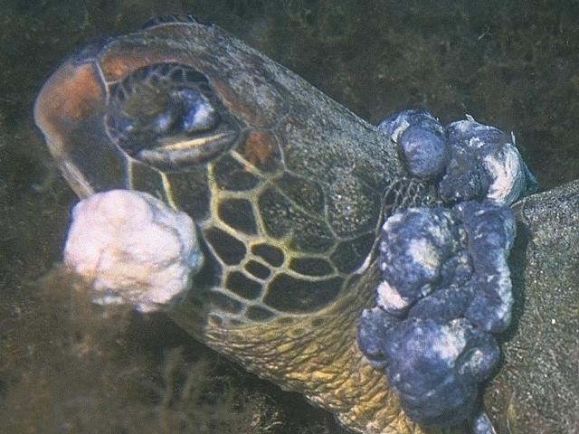
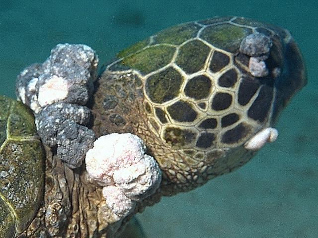 Right side, 1992 Turtle 22: 4 Spot (58K JPEG)
This is 4 Spot's right side. Tumors do not cover the eye completely yet. The tumor at the right side of the mouth is larger than on the left. The right side of the jaw is swollen and bulging, possibly from growth that we cannot see.
 1995 Turtle 38 [unnamed] (70K JPEG)
This was the weakest, most emaciated turtle we have ever seen. This was a small turtle, perhaps 40 cm carapace length. It had a blackened, concave plastron, and was covered in algae.
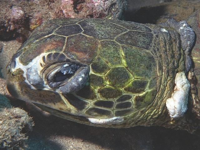
 1993 Turtle 11 [unnamed] (70K JPEG)
In 1993, this turtle showed no external signs of tumors. By 1994, there had been a dramatic outbreak. In 1995, although the size and number of tumors had not increased significantly, the animal was emaciated and monofilament fishing line had gotten tangled in its tumors.
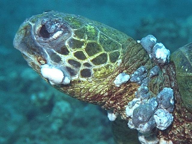
 1995 Turtle 39 [unnamed] (59K JPEG)
In 1995, we saw this turtle for the first time. It stayed close to the Graveyard, a shallow area with plentiful food and the place at which we see the worst cases. The tumors around the neck were dark and shrunken, but the mouth and eye tumors were whitish. In our experience, this indicates that these tumors are still growing.
 1995 Turtle 40 [unnamed] (55K JPEG)
This is another turtle that we saw for the first time in 1995. Like Turtle 39, this turtle stayed near the Graveyard. This turtle was so badly burdened with tumors that swimming took enormous effort, usually consisting of swimming a short distance, surfacing for air, and then settling on the bottom to rest before continuing.
If there are specific images you would like to see in the Turtle Trax pathology pages, let us know. Please send comments or suggestions to webmaster@turtles.org.
 Sickbay
 Turtle Trax Home Page